A wound becoming smaller, a participant liking a treatment, and an investigator seeing better-looking skin are three different kinds of evidence. Each can matter, but none substitutes automatically for the others. Copper-peptide research illustrates this distinction unusually well: laboratory experiments examine cellular activity, a diabetic-ulcer study measures wound closure, and a post-laser study compares both visible outcomes and participant assessments.
The central question is not whether one positive result makes copper peptides broadly effective. It is what was measured, against which comparator, in which tissue, and over what period. This analysis uses an author-posted primary full-text OCR of the ulcer paper, its indexed abstract, and indexed primary abstracts for the laser and cell experiments. The original ulcer tables and figures were not visually verified. The aim is to preserve the clinical finding while keeping its meaning narrower than an ingredient-wide promise.
Download chart data (CSV)Download chart (PNG)
Study facts at a glance
Historical publication: The ulcer paper was published in 1994, not in the later year suggested by its PubMed indexing. [1]
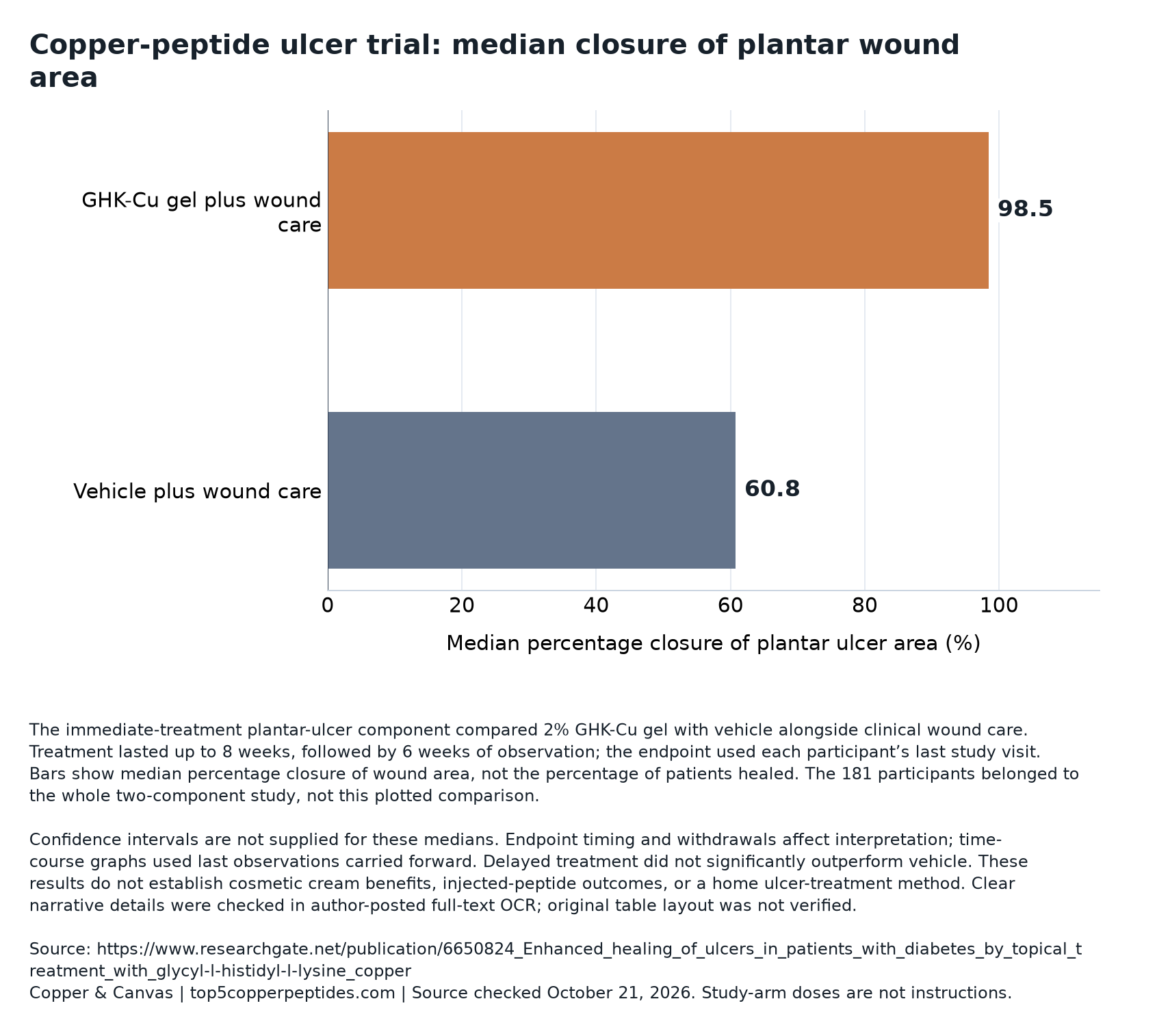
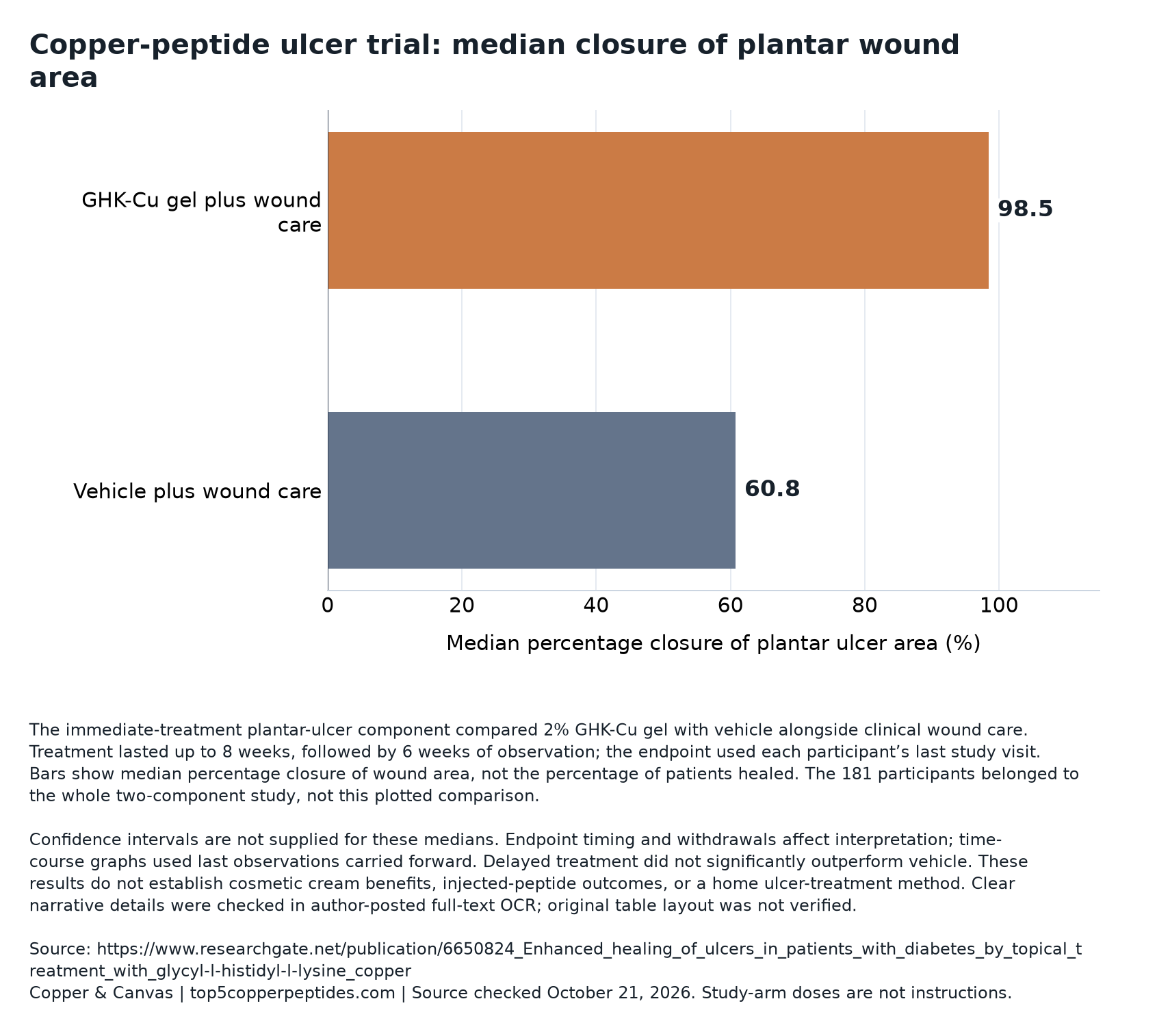
Main area result: The immediate-treatment plantar-ulcer comparison reported median area closure of 98.5% with active gel versus 60.8% with vehicle—not percentages of participants completely healed. [2] [1]
Larger-ulcer finding: The abstract reported median area closure of 89.2% versus −10.3% in its larger-ulcer subgroup. [2]
Infection finding: Reported infections were 7% versus 34%, specifically in plantar ulcers treated immediately after debridement. [2]
Different kinds of benefit: The laser study’s positive participant questionnaire result was distinct from its nonsignificant between-group objective comparisons. [3]
Related reading: What copper peptides can—and cannot—tell you · CoreAge Rx Bounce Back review
The experiment tested an addition to organized wound care
The randomized, multicenter, evaluator-blinded trial enrolled 181 people, averaging 60 years of age, with diabetic neuropathic ulcers. All received standardized debridement, pressure-relieving footwear, and education. Immediate treatment compared topical 2% GHK-Cu gel with vehicle for up to eight weeks, followed by six weeks of observation. A delayed component used four weeks of vehicle before eight further weeks of vehicle, 2%, or 4% gel.
That design asks an incremental question: does adding the active ingredient improve outcomes beyond the care and gel base shared by the groups? It does not compare copper peptide with doing nothing. A vehicle is the formulation without the tested active ingredient; using it helps separate ingredient effects from effects associated with the formulation and treatment process. Randomization supports a causal comparison, while blinded evaluation reduces the opportunity for expectations to influence measurements.
Interpretation also depends on keeping the experimental unit straight. A whole-study enrollment figure describes the research program, not necessarily the denominator for one anatomical subgroup or treatment-timing comparison. Combining those levels would make a restricted result appear more broadly demonstrated than it was. These historical concentrations identify study arms; they are not instructions for treating a wound or selecting a cosmetic percentage.
Area closure measures a continuous change, not a head count
Percentage area closure describes how much of the starting open area is no longer open. In a simplified calculation, subtract the later area from the starting area, divide by the starting area, and multiply by 100. The result is a continuous measurement: a wound can improve substantially without reaching complete closure. A median summarizes the middle of a distribution of such measurements; it does not count how many participants crossed a healing threshold.
For illustration, imagine three wounds with area reductions of 40%, 90%, and 100%. Their median reduction is 90%, although only one wound has reached complete area closure. This invented example is not a reconstruction of the trial. It shows why converting a median percentage into a percentage of healed patients changes the endpoint. The trial separately assessed near-complete closure categorically, but ambiguous OCR tables prevent reliable publication of those arm-specific proportions here.
The distinction matters beyond wound research. Whenever a headline contains a percentage, identify its denominator and its operation: percentage change within a wound, percentage of people achieving an outcome, or difference between groups. Our guide to reading GHK-Cu evidence provides a broader framework for keeping ingredient activity, measured outcomes, and product claims separate.
| Measurement | What it describes | What it does not establish |
|---|---|---|
| Percentage area closure | Change relative to a wound’s starting area | The percentage of participants completely healed |
| Categorical closure | Whether an ulcer crosses a specified closure threshold | The full distribution of improvement below that threshold |
| Normalized edge-advance rate | Movement of the wound margin per unit of time | How many times more participants healed |
Compare the options in top 5 copper peptide creams & serums or read Bounce Back versus The Ordinary copper peptides.
Negative closure means enlargement, with an asymmetric scale
A negative area-closure percentage is mathematically coherent. In an illustrative wound starting at 200 square millimeters, enlargement to 220 square millimeters produces −10% closure. Nothing has become a negative number of healed patients; the wound has become larger. Conversely, reduction to zero open area produces 100% closure. Improvement has a ceiling, while proportional deterioration can extend much farther in the negative direction.
That asymmetry helps explain why means and medians may tell different stories. Consider five hypothetical closure values: −200%, 60%, 80%, 90%, and 100%. The median is 80%, but the mean is 26%. Neither calculation is wrong. The median conveys the middle response; the mean is pulled down by major worsening. A middle response therefore should not be read as a complete account of outcomes across participants.
The abstract’s negative larger-ulcer value and the body OCR’s enlargement figure differ by 0.1 percentage point. The threshold is also described as greater than 100 square millimeters in the abstract and at least 100 in the body. These unresolved details favor referring to study-defined larger ulcers around that threshold. They do not erase the reported direction, but they prevent pretending that transcription-level precision has been independently confirmed.
A faster edge-advance rate is a different endpoint again
The ulcer abstract described closure as three times faster; the full report identifies a normalized linear edge-advance measure in millimeters per day. Think of this as estimating how quickly the wound boundary moves inward while accounting for wound geometry. It is not simply the number of square millimeters lost each day, and it is not a ratio of the numbers of people healed.
Geometry makes that distinction useful. As an illustrative model, suppose two circular wounds have different radii but their boundaries both move inward by the same distance. The larger circle can lose more area even though its edge moves at the same speed. Comparing raw area losses alone could therefore confuse starting size with biological progress. A normalized rate attempts to answer a more comparable question across differently sized wounds.
A rate ratio also needs an absolute scale. A large ratio can arise when the comparator rate is small, and it does not tell readers how long every wound will take to close. Nor does an average rate guarantee steady progress within an individual wound. To understand a treatment fully, readers need the rate definition alongside closure distributions and clinically meaningful outcomes, rather than letting one striking multiplier stand in for all three.
Treatment timing generated a question, not a proven mechanism
Delayed treatment did not show significant improvement over vehicle. That contrasts with the immediate-treatment finding, but it does not establish the biological reason for the difference. Timing can change the tissue environment, the stage of healing, and the meaning of a baseline comparison. A study can detect a pattern without measuring the intermediate steps needed to explain it.
The methodological issue is especially important when an early phase is used as a reference for a later phase. Such a comparison implicitly assumes that the untreated trajectory would remain sufficiently comparable over time. If healing naturally changes speed after the initial intervention, a later rate cannot be interpreted simply by subtracting the earlier one. A slowing trajectory might reflect disease dynamics, treatment effects, or both.
A stronger mechanistic investigation would separate these possibilities prospectively, define timing comparisons in advance, and measure proposed intermediate processes. That is an interpretation of what further evidence would be useful, not a claim that such measurements were performed. Likewise, a nonsignificant delayed comparison means that superiority was not demonstrated under those conditions; it does not mean the wounds remained unchanged. Higher concentration also cannot be assumed to compensate for a different treatment context.
The last visit is not one shared follow-up date
Weekly wound tracings underwent planimetry. The endpoint used each participant’s last visit, including closure or discontinuation; longitudinal graphs carried the last value forward. All enrollees entered the analyses, including four with only baseline evaluation. Infection unresponsive after a week of treatment triggered withdrawal.
These choices preserve records, but they do not make observation complete. An endpoint measured when a participant leaves is not necessarily an endpoint measured at the same scheduled week for everyone. Someone leaving after successful closure and someone leaving after deterioration contribute observations with different clinical meanings. A statement about results after a fixed period would require everyone’s outcome to be evaluated at that period, or an explicit method for estimating it.
Carrying a last observation forward makes a further assumption: the recorded state stands in for later unobserved states. That can be a practical plotting convention without being a biological truth. A closed wound might reopen, and a worsening wound might later improve or deteriorate further. The direction of bias cannot be assigned confidently without additional information. This is why inclusion in an analysis and completeness of follow-up are separate properties, not interchangeable badges of reliability.
Infections and overall adverse events require separate readings
The report found no significant difference in overall adverse-event rates. Exact event counts could not be reliably verified from the available transcription; confidence intervals were not supplied for the cited comparisons. Its lower infection result belongs to the immediate plantar comparison, not the entire trial. A favorable local outcome and an inconclusive overall safety comparison can coexist.
An arithmetic illustration clarifies the infection contrast: subtracting the reported group percentages yields a 27-percentage-point difference. That is not the same as a 27% relative reduction. More importantly, a difference calculated from subgroup percentages is not automatically a dependable estimate for a different patient population. Without verified denominators and uncertainty intervals, converting it into a treatment guarantee or a number needed to treat would imply unsupported precision.
Safety interpretation also depends on who was eligible, what was monitored, and how long observation lasted. A comparison lacking statistical significance does not prove equal safety; it may be compatible with a range of differences. Conversely, the infection finding deserves recognition rather than being dismissed because it is restricted. The disciplined reading is to retain both facts: a specific favorable signal and unresolved uncertainty about the broader adverse-event profile.
Participant experience and investigator assessment can diverge
In a separate randomized post-CO2 circumoral resurfacing study, 13 patients completed regimens with or without GHK-Cu. At 12 weeks, objective wrinkle and overall appearance comparisons did not differ significantly between groups; erythema assessments were also nonsignificant. A participant questionnaire favored GHK-Cu for overall skin quality or satisfaction, with P = .04. Enrollment, arm sizes, effect sizes, confidence intervals, and adverse-event counts were unreported in the abstract.
There is no contradiction in these outcomes. A participant questionnaire and an investigator assessment measure related but nonidentical experiences. Satisfaction can be valuable in its own right without establishing a visible structural change. Equally, a small objective comparison that does not reach significance does not establish equivalence. The useful question is whether the outcome matches the claim being made, not which endpoint sounds most favorable.
Both groups improving after a shared procedure would not, by itself, identify an added ingredient effect. The causal comparison remains the difference between regimens. Our closer look at the laser-resurfacing trial develops that distinction. Across the two clinical settings, wound tracing, visible skin assessment, and participant experience should remain separate evidence categories rather than being merged into a general score for copper peptides.
| Research setting | Measured signal | Appropriate boundary |
|---|---|---|
| Diabetic-ulcer trial | Wound closure and ulcer-related events | Clinical wound outcomes within a structured care protocol |
| Post-laser trial | Objective skin assessments and participant questionnaire | Separate visible outcomes from participant-reported experience |
| Fibroblast experiments | Collagen synthesis and remodeling-related signals | Cellular plausibility, not demonstrated cosmetic benefit |
Cell experiments describe remodeling, not a finished cosmetic result
One fibroblast-culture experiment reported increased collagen synthesis without increased cell number. Another reported increased MMP-2 and its messenger RNA, alongside increased TIMP-1 and TIMP-2 secretion. Copper ions reproduced the MMP-2 effect, whereas GHK alone did not. These findings concern cells and their surroundings, not treated faces.
Collagen is part of the extracellular matrix, the supporting material around cells. MMPs are enzymes involved in matrix breakdown and remodeling; TIMPs inhibit metalloproteinases. Observing changes in both helps resist an overly simple narrative in which more signaling must mean more accumulated collagen. Tissue remodeling involves production, removal, organization, and regulation. A cellular increase in one component does not specify the final architecture or appearance of living skin.
Translation therefore requires several additional tests: whether a formulation delivers the relevant material to the relevant tissue, whether the cellular effect occurs there, and whether it produces a meaningful human outcome without unacceptable harms. Laboratory concentrations do not answer those questions or supply product-use instructions. The copper-peptide overview and percentage-comparison guide are useful companions for distinguishing chemical identity from demonstrated delivery and benefit.
Product decisions need a separate documentary record
ProCyte affiliations, the named study product, and an acknowledged development team document commercial involvement, but not a formal funding statement. The readable narrative supports the trial’s main interpretation; scrambled tables do not justify publishing unverified arm counts. Confidence intervals for the central outcome estimates are unavailable. These are different transparency questions and should not be collapsed into either blanket trust or blanket dismissal.
Editorial interpretation: a product decision has at least two evidence tracks. One asks whether the exact formulation, route, and intended outcome have been tested. The other asks what a seller’s documents actually promise about payment, cancellation, fulfillment, and returns. A clinical paper cannot answer the second track. Readers can consult the CoreAge Rx review or Likeness Rx review to check documentary terms, without treating either link as evidence of studied-product availability or clinical outcomes.
Likewise, the cream-versus-serum comparison addresses a formulation question, not proof of equivalence to a wound gel. Clinical results do not transfer automatically to providers, compounded preparations, oral products, systemic injections, or retail facial formulas. The scientifically useful next step is matched evidence: the intended population, finished formulation, comparator, and outcome studied together, with transparent follow-up and uncertainty.
What this evidence supports
The ulcer research supports a bounded clinical signal, not a universal copper-peptide benefit. Its strongest interpretive lesson is to keep measurement types separate: wound-area reduction, normalized closure speed, infection events, visible skin assessment, and participant satisfaction each answer different questions. The clinical comparison deserves attention within its treatment setting, while uncertain follow-up assumptions and unverified table details limit precision. For facial skincare, biological plausibility remains a reason to conduct matched clinical studies—not a shortcut around them.
Research access and limitations
The ulcer analysis uses an author-posted primary full-text OCR and the indexed abstract. Original table and figure layouts were not verified; ambiguous arm counts and categorical rates are therefore omitted. The larger-ulcer threshold and a 0.1-percentage-point enlargement discrepancy remain unresolved. The laser and cell studies were available as indexed primary abstracts only; full protocols and supplements were not examined. No current regulatory status, formal trial funding arrangement, or equivalence of commercial formulations is inferred.
Questions about the evidence
Does a high median closure percentage mean almost everyone healed?
No. A median is the middle wound-level measurement, not a count of people reaching complete healing. A categorical endpoint answers the latter question. Even near-complete area reduction should not silently become complete healing, because changing the threshold changes which outcomes qualify. [2] [1]
Why not report only the strongest subgroup result?
A subgroup can identify a clinically interesting pattern, but it answers a narrower question. Its credibility depends on how the group was defined, how many comparisons were made, and the precision of its estimate. It should be presented alongside the broader design rather than substituted for the entire experiment. [2] [1]
Is participant satisfaction less scientific than an investigator assessment?
Not inherently. A well-designed participant questionnaire can measure an important experience. The limitation is matching: satisfaction evidence supports an experience-related conclusion, while a claim about visible wrinkles or wound closure needs a corresponding assessment. Neither endpoint should be relabeled as the other. [3]
Would more copper peptide necessarily produce a larger benefit?
No dose rule follows from these studies. Concentration, formulation, tissue access, timing, and outcome are distinct variables. A laboratory response or a historical gel arm does not establish a useful concentration for a different finished product, particularly across different routes or tissue conditions. [1] [4] [5]
What evidence would most improve confidence in a cosmetic claim?
A prospective comparison of the finished cosmetic formula against an appropriate control, with a clearly defined visible outcome, adequate enrollment, transparent attrition, adverse-event reporting, and confidence intervals would directly address that claim. Cell activity and wound research can motivate such testing, but cannot replace it. [3] [1] [4] [5]
Sources and verification
Official pages establish product facts and provider claims. Clinical and regulatory sources answer separate evidence questions. Article checked October 21, 2026; individual source dates appear below. No medical reviewer is credited for this article.
- Mulder et al., 1994: GHK-Cu in diabetic neuropathic ulcers (author-posted full text) ↗ · Checked October 21, 2026 · Author-posted primary full-text OCR; original table and figure layout not verified
- Enhanced healing of ulcers in patients with diabetes by topical treatment with glycyl-l-histidyl-l-lysine copper. ↗ · Checked October 21, 2026 · Indexed primary study abstract
- Effects of topical copper tripeptide complex on CO2 laser-resurfaced skin. ↗ · Checked October 21, 2026 · Indexed primary study abstract
- Stimulation of collagen synthesis in fibroblast cultures by the tripeptide-copper complex glycyl-L-histidyl-L-lysine-Cu2+. ↗ · Checked October 21, 2026 · Indexed primary laboratory-study abstract
- The tripeptide-copper complex glycyl-L-histidyl-L-lysine-Cu2+ stimulates matrix metalloproteinase-2 expression by fibroblast cultures. ↗ · Checked October 21, 2026 · Indexed primary laboratory-study abstract